| SKU-Pack Size | Availability | Size | Price | |
| EAB11650-30UL | In Stock | 30ul | €115.70 | |
| EAB11650-100UL | In Stock | 100ul | €258.70 | |
| EAB11650-200UL | In Stock | 200ul | €440.70 |
| Please select the country you are in to find your local distributor. |
| Dianost | Phone£º+49 (0) 40 65472-053 | |
| Bullenkoppel 11£¬ 23896 Poggensee, | E-mail£ºola@olasciences.com | |
| Germany | Germany | Web£ºwww.olasciences.com |
| Product Information | |
| Applications | WB, ELISA |
| Species Reactivity | Human, Mouse, Rat |
| Host / Isotype | Rabbit IgG |
| Clonality | Polyclonal |
| Applications Dilutions | WB=1:500-2000 |
| MW (kDa) | 112 |
| Conjugate | Unconjugate |
| Specificity | OPA1 Rabbit Polyclonal Antibody detects endogenous levels of OPA1 protein. |
| Purification | Affinity purification |
| Concentration | 1mg/ml |
| Format | Liquid |
| Formulation | In PBS, pH 7.4, containing 0.02% sodium azide,0.5% BSA and 50% glycerol. |
| Shipping | Gel Pack |
| Storage | Store at -20¡ãC least 1 year from the date of shipment. avoid repeated freeze/thaw cycles. Aliquots may be stored at +4¡ãC for 1-2 weeks. |
| Research Use | For Research Use Only. Not Intended for Diagnostic or Therapeutic Use. |
| Application Key WB-Western Blot IP-Immunoprecipitation IHC-Immunohistochemistry IF-Immunofluorescence ICC-Immunocytochemistry FC-Flow Cytometry |
|
| Reactivity Key H-Human M-Mouse R-Rat Mk-Monkey B-Bovine Pg-Pig Hm-Hamster Dg-Dog C-Chicken X-Xenopus Z-Zebrafish Hr-Horse All-All Species Expected |
|
| Product Bioinformatics | |
| Synonym(s) | Optic atrophy 1; NPG; NTG; MGM1; BERHS; largeG; MTDPS14; OPA1 |
| Gene Aliases | OPA1 |
| UniProt ID | |
| Entrez Gene ID | |
| Product Description | |
OPA1 is a nuclear-encoded mitochondrial protein with similarity to dynamin-related GTPases. OPA1 plays a crucial role in mitochondrial dynamics, particularly in regulating mitochondrial fusion and maintaining mitochondrial morphology. This function is vital for cellular energy production and overall cell health, helping ensure mitochondria can efficiently adapt to changes in energy demand and stress conditions. Defects in OPA1 are associated with optic atrophy type 1, leading to vision loss, and OPA1 is predominantly expressed in the retina, although also found in the brain, testis, heart, and skeletal muscles. OPA1′s ability to interact with other proteins involved in mitochondrial function further underscores OPA1′s importance in cellular metabolism and the pathophysiology of mitochondrial diseases.
|
| Product Image Gallery | |
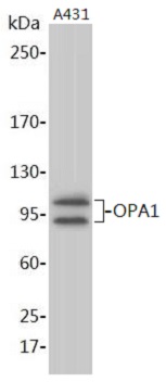
![]() Western blot analysis of extracts on A431 cells, using OPA1 Rabbit Polyclonal Antibody (EAB11650) at 1:1000 dilution. Secondary antibody Goat Anti-Rabbit IgG (H&L)-HRP (EAB21002) at 1:5000 dilution.
Predicted band size: 112 kDa; Observed band size: 105, 80-95 kDa |
| Specific Protocols | |