

www.ebiocell.com
| Host / Isotype | Clonality | Applications | MW (kDa) | Reactivity |
| Rabbit IgG | Polyclonal | WB, IHC-P, IF, ELISA | 52 | Human, Mouse, Rat |
| Applications Dilutions | |
| The application notes include recommended starting dilutions;optimal dilutions/concentrations should be detemined by the end user. | |
| WB=1:500-2000 IHC-P=1:50-300 IF=1:50-300 |
|
| Application Key | WB-Western Blot IP-Immunoprecipitation IHC-Immunohistochemistry IHC-P-Immunohistochemistry-Paraffin |
| Reactivity Key | H-Human M-Mouse R-Rat Mk-Monkey B-Bovine Pg-Pig Hm-Hamster Dg-Dog C-Chicken X-Xenopus Z-Zebrafish |
| Product Information | |
| Conjugate | Unconjugate |
| Specificity | Phospho-Smad2 (Ser467) Rabbit Polyclonal Antibody detects endogenous levels of Smad2 only when phosphorylated at Ser467. |
| Purification | Affinity purification |
| Concentration | 1mg/ml |
| Format | Liquid |
| Formulation | In PBS, pH 7.4, containing 0.02% sodium azide,0.5% BSA and 50% glycerol. |
| Shipping | Gel Pack |
| Storage | Store at -20°C least 1 year from the date of shipment. avoid repeated freeze/thaw cycles. Aliquots may be stored at +4°C for 1-2 weeks. |
| UniProt ID | |
| Entrez Gene ID | |
| Product Description | |
Smad2 also known as MADR2 is a critical intracellular mediator in the Transforming Growth Factor-beta (TGF-β) signaling pathway. Smad2 plays a crucial role in the transforming growth factor-beta (TGF-β) signaling pathway, which is essential for regulating cellular processes such as proliferation, differentiation, and apoptosis. Smad2 is primarily located in the cytoplasm and nucleus, where Smad2 translocates upon phosphorylation in response to TGF-β signaling. This localization enables Smad2 to act as a transcription factor, regulating the expression of target genes that mediate the cellular response to TGF-β. Smad2′s ability to shuttle between the cytoplasm and nucleus underscores Smad2′s importance in cellular signaling and gene regulation, making Smad2 a key player in developmental processes and tissue homeostasis. Smad2 interacts with other Smad proteins, such as Smad3 and Smad4, to form complexes that facilitate the transcription of TGF-β target genes, further emphasizing Smad2′s role in the intricate network of cellular signaling pathways. |
| Product Image Gallery | |
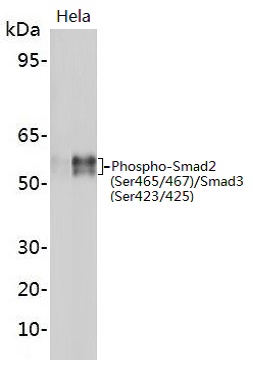 Western blot analysis of extracts on HepG2 cells treated with PMA, using Phospho-Smad2 (Ser467) Rabbit Polyclonal Antibody (EAB13989) at 1:1000 dilution. The lane on the left is blocked with the phospho peptide.Predicted band size: 52 kDa; Observed band size: 58 kDa
|
