

| Host / Isotype | Clonality | Applications | MW (kDa) | Reactivity |
| Mouse IgG1 | Monoclonal | WB, IF/ICC, FC, ELISA | 82 | Human, Mouse, Rat |
| Application Key | WB-Western Blot IP-Immunoprecipitation IHC-Immunohistochemistry IHC-P-Immunohistochemistry-Paraffin |
| Reactivity Key | H-Human M-Mouse R-Rat Mk-Monkey B-Bovine Pg-Pig Hm-Hamster Dg-Dog C-Chicken X-Xenopus Z-Zebrafish |
| Applications Dilutions | |
| The application notes include recommended starting dilutions;optimal dilutions/concentrations should be detemined by the end user. | |
| WB(Westem Blotting) IHC-P(Immunohistochemistry-Paraffin) IF/ICC(Immunofluorescence/Immunocytochemistry) ELISA(Enzyme-linked Immunosorbent Assay) |
WB=1:500-2000 IF/ICC=1:100-500 FC=1:50-200 |
| Product Information | |
| Conjugate | Unconjugate |
| Specificity | DRP1 Mouse Monoclonal Antibody detects endogenous levels of DRP1 protein. |
| Purification | Affinity purification |
| Concentration | 1mg/ml |
| Format | Liquid |
| Formulation | In PBS, pH 7.4, containing 0.02% sodium azide,0.5% BSA and 50% glycerol. |
| Shipping | Gel Pack |
| Storage | Store at -20°C least 1 year from the date of shipment. avoid repeated freeze/thaw cycles. Aliquots may be stored at +4°C for 1-2 weeks. |
| UniProt ID | |
| Entrez Gene ID | |
| Product Description | |
DRP1 also known as Dynamin-like protein 1, Dlp1, DVLP, DNM1L is a member of the dynamin superfamily of GTPases. DRP1 plays a crucial role in mediating outer mitochondrial membrane fission in mammalian cells, a process essential for mitochondrial dynamics and cellular health. DRP1 is characterized by an N-terminal tripartite GTP-binding domain, which is a hallmark of the Dynamin superfamily of GTPases. Structurally, DRP1 exists as a T-shaped dimer composed of a head, leg, and stalk, and GTP binding induces a conformational change that generates the mechanical force necessary for membrane constriction. DRP1 is ubiquitously expressed, with particularly high levels in skeletal muscle, heart, kidney, and brain tissues, and localizes to the perinuclear region within cells. In mouse brain, DRP1 shows high expression in the cerebellum, especially in Purkinje cells, highlighting DRP1′s importance in neuronal function. During apoptosis, DRP1 translocates from the cytosol to mitochondria, localizing to sites of organe. |
| Product Image Gallery | |
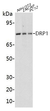 Western blot analysis of extracts on NIH/3T3, LNCAP, PC12 cells, using DRP1 Mouse Monoclonal Antibody (EAB21209) at 1:1000 dilution. Secondary antibody Goat Anti-Mouse IgG (H&L)-HRP (EAB21001) at 1:5000 dilution.
|
