
| Host / Isotype | Clonality | Applications | MW (kDa) | Reactivity |
| Rabbit IgG | Monoclonal | WB, IHC-P, IF/ICC, FC | 67 | Human, Mouse, Rat |
| Application Key | WB-Western Blot IP-Immunoprecipitation IHC-Immunohistochemistry IHC-P-Immunohistochemistry-Paraffin |
| Reactivity Key | H-Human M-Mouse R-Rat Mk-Monkey B-Bovine Pg-Pig Hm-Hamster Dg-Dog C-Chicken X-Xenopus Z-Zebrafish |
| Applications Dilutions | |
| The application notes include recommended starting dilutions;optimal dilutions/concentrations should be detemined by the end user. | |
| WB(Westem Blotting) IHC-P(Immunohistochemistry-Paraffin) IF/ICC(Immunofluorescence/Immunocytochemistry) ELISA(Enzyme-linked Immunosorbent Assay) |
WB=1:500-2000 IHC-P=1:50-200 IF/ICC=1:50-200 IP=1:10-100 |
| Product Information | |
| Conjugate | Unconjugate |
| Specificity | TAK1 Rabbit Monoclonal Antibody detects endogenous levels of TAK1 protein. |
| Purification | Affinity purification |
| Concentration | 1mg/ml |
| Format | Liquid |
| Formulation | In PBS, pH 7.4, containing 0.02% sodium azide,0.5% BSA and 50% glycerol. |
| Shipping | Gel Pack |
| Storage | Store at -20°C least 1 year from the date of shipment. avoid repeated freeze/thaw cycles. Aliquots may be stored at +4°C for 1-2 weeks. |
| UniProt ID | |
| Entrez Gene ID | |
| Product Description | |
TAK1 also known as is TGF1a, MAP3K7 is a member of the serine/threonine protein kinase family. TAK1 plays a pivotal role in mediating cellular responses to cytokines like TGFβ and interleukin-1 (IL-1) and controls a variety of cell functions including transcription regulation and apoptosis. In response to IL-1, TAK1 forms a kinase complex including TRAF6, MAP3K7P1/TAB1 and MAP3K7P2/TAB2 activating downstream NF-κB and MAP kinase signaling pathways essential for inflammation, immune responses, cell survival, and apoptosis. Dysregulation of Tak1 function is associated with various pathological conditions, including cancer and inflammatory diseases, making Tak1 a critical target for biomedical research. |
| Product Image Gallery | |
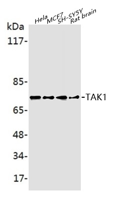 Western blot analysis of extracts on Hela, MCF7, SH-SY5Y, Rat brain cells, using TAK1 Rabbit Monoclonal Antibody (EAB22824) at 1:1000 dilution. Secondary antibody Goat Anti-Rabbit IgG (H&L)-HRP (EAB21002) at 1:5000 dilution.
Predicted band size: 67 kDa; Observed band size: 75 kDa |
